
Download
G S H = Y − 0.00314 0.0314 × D F B T × V u MDA concentration = Y × 100 × V t 1.560000 × W t × V u , C A T = δ 0 D E × volume of sample m L × protein m g , R = 5.74 × 10 − 4 A C o , R = 5.74 × 10 − 4 × A C o , 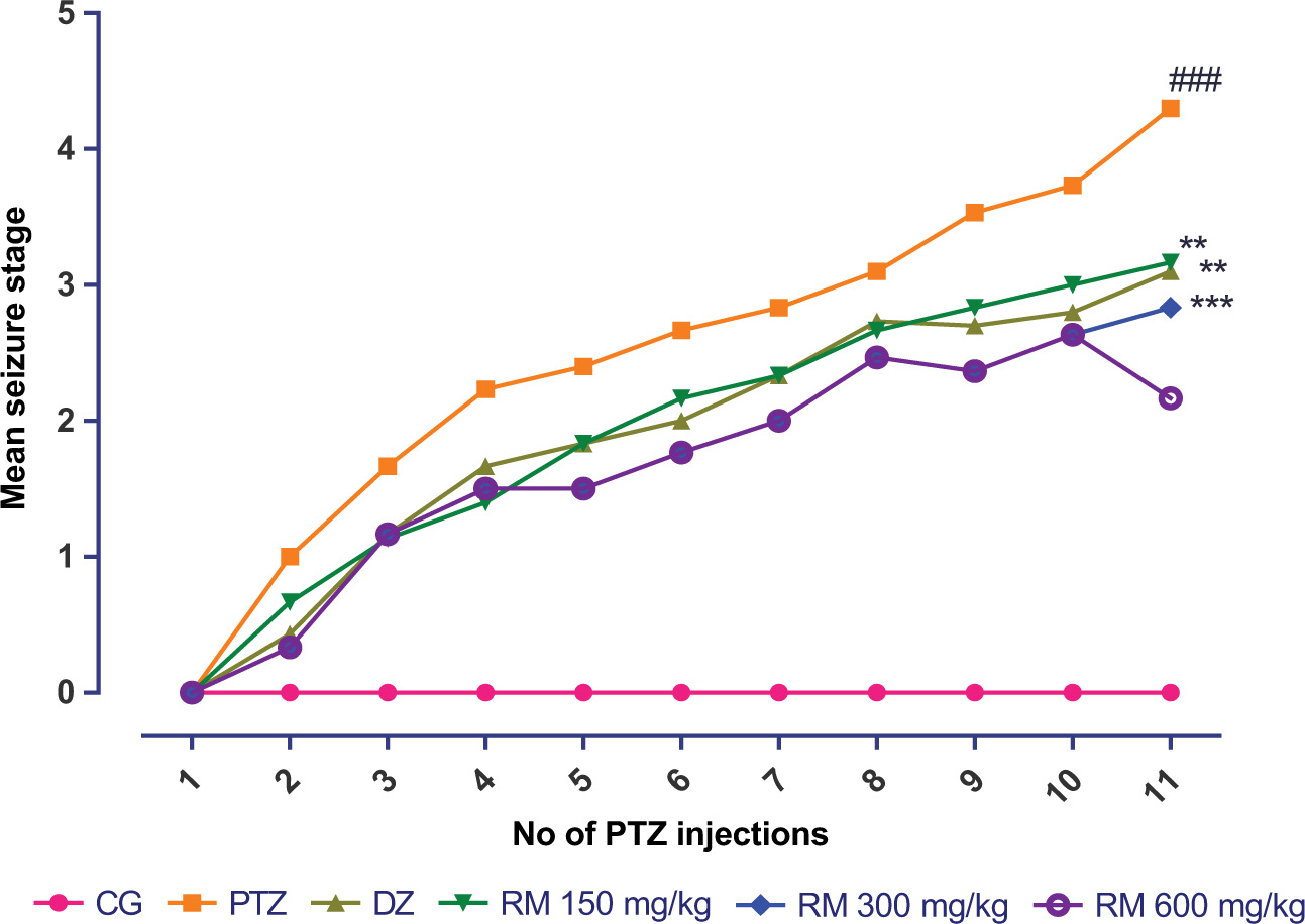
PAPER
Rosa moschata leaf extract mitigates seizures, oxidative stress, and neurotransmitter imbalance in pentylenetetrazol-induced mice
Humidah Alanazi1, Humaira Gul2, Maryam Farrukh2, Uzma Saleem3*, Areej M. Alsolami4, Arooj Anwer2, Wafa S. Al-Thubiani5, Ibrahim Aljaezi6, Nouf Abdullah Alharbi7, Alaa K. Khojah8, Ali G. Alkhathami9, Aishah Albalawi10, Nada Alhazmi11,12, Muhammad Ajmal Shah13,14,15*, Agustina Lulustyaningati Nurul Aminin14, Puteri Amelia15, Ana Sanches Silva16*, Norah K. Algarzae17*
1Department of Biochemistry, College of Science, King Saud University, Riyadh, Saudi Arabia;
2Department of Pharmacology, Faculty of Pharmaceutical Sciences, Government College University, Faisalabad, Pakistan;
3Punjab University College of Pharmacy, University of Punjab, Lahore, Pakistan;
4Department of Biology, College of Science, Jouf University, P.O. Box 2014, Sakaka, Al-Jouf, Saudi Arabia;
5Department of Biology, Faculty of Science, Umm Al-Qura University, Mecca, Saudi Arabia;
6Department of Pharmacology, College of Pharmacy, Najran University, Najran, Saudi Arabia;
7Department of Basic Health Sciences, College of Applied Medical Sciences, Qassim University, Buraydah, Saudi Arabia;
8Department of Pharmaceutical Sciences, College of Pharmacy, Umm Al-Qura University, Makkah, Saudi Arabia;
9Department of Clinical Laboratory Sciences, College of Applied Medical Sciences, King Khalid University, Abha, Saudi Arabia;
10Department of Biology, Faculty of Science, University of Tabuk, Tabuk, Saudi Arabia;
11Department of Basic Sciences, College of Science and Health Professions, King Saud bin Abdulaziz University for Health Sciences, Jeddah, Saudi Arabia;
12King Abdullah International Medical Research Center, Jeddah, Saudi Arabia;
13Department of Pharmacy, Hazara University, Mansehra, Pakistan;
14Department of Chemistry, Faculty of Science & Mathematics, Diponegoro University, Semarang, Indonesia;
15Department of Pharmacy, Faculty of Health Sciences, Universitas Islam Negeri Syarif Hidayatullah, Jakarta, Indonesia;
16Faculty of Pharmacy, University of Coimbra, Polo III, Azinhaga de Sta Comba, Coimbra, Portugal;
17Department of Physiology, College of Medicine, King Saud University, Riyadh, Saudi Arabia
Abstract
The crippling neurological state, known as epilepsy, is typified by frequent spontaneous seizures and linked to oxidative stress-driven neuronal dysfunction; it presents a significant global health challenge. This study investigates the traditional neuroprotective claims for Rosa moschata by rigorously evaluating the neuroprotective and anticonvulsant potential of its ethanolic leaf extract in a pentylenetetrazol (PTZ, 35 mg/kg)-induced kindling epilepsy model of mice. Oral administration of Rosa moschata leaf extract (RMLE) at different doses (150, 300, and 600 mg/kg) for 3 weeks beside PTZ challenge demonstrated significant, dose-dependent protection. RMLE markedly improved neurobehavioral outcomes, enhancing performance in various behavioral tests, indicating improved cognition, reduced anxiety, and better motor coordination. Crucially, RMLE restored critical neurotransmitter balance, significantly elevating brain levels of serotonin, dopamine, and noradrenaline, but suppressing acetylcholinesterase performance. The extract exhibited potent antioxidant efficacy, substantially increasing superoxide dismutase, catalase, and reduced glutathione levels, while effectively decreasing malondialdehyde and nitrite concentrations, thereby countering PTZ-induced oxidative stress. Histopathological examination of the brain revealed a compelling
dose-dependent reduction in neurofibrillary tangles and plaques. Comprehensive safety assessment through liver and kidney function tests (LFT and RFT), complete blood count, and histopathology of the brain, heart, liver, and kidney confirmed no significant adverse effects, highlighting a favorable safety profile. These findings provide robust experimental validation: RMLE possesses significant anticonvulsant activity mediated through multi-faceted mechanisms, including neurotransmitter modulation, potent antioxidant action, mitigation of neuronal pathology, and absence of detectable toxicity, thereby strongly supporting its traditional use and identifying it as a promising candidate for epilepsy management.
Key words: dopamine, epilepsy, noradrenaline, oxidative stress biomarkers, serotonin
*Corresponding authors: Uzma Saleem, Punjab University College of Pharmacy, University of Punjab, Lahore, Pakistan. Email: [email protected]; Muhmmad Ajmal Shah, Department of Pharmacy, Hazara University, Mansehra, Pakistan. Email: [email protected]; Ana Sanches Silva, Faculty of Pharmacy, University of Coimbra, Polo III, Azinhaga de Sta Comba, Coimbra, Portugal. Email: [email protected]; Norah K. Algarzae, Department of Physiology, College of Medicine, King Saud University, Riyadh, Saudi Arabia. Email: [email protected]
Academic Editor: Prof. Tommaso Beccari, University of Perugia, Italy
Received: 27 June 2025; Accepted: 25 August 2025; Published: 17 October 2025
© 2025 Codon Publications
This is an Open Access article distributed under the terms of the Creative Commons Attribution-NonCommercial-ShareAlike 4.0 International (CC BY-NC-SA 4.0). License (http://creativecommons.org/licenses/by-nc-sa/4.0/)
Introduction
The peripheral nerves, spinal cord, and brain are affected by a wide range of sensory system abnormalities known as neurological diseases (Fischer et al., 2015). They are characterized by several sets of enzymes and anatomical defects. According to different studies, continual neuronal deterioration and destruction of the sensory system are due to neuro-inflammation, proteostasis strains, oxidative pressure, and apoptosis (Narne et al., 2017). Neuronal loss because of oxidative and excitotoxicity mechanisms has been hypothesized to play a major role (Sevindik et al., 2021).
Epilepsy is a condition described by two or more unprovoked seizures, which are not immediately linked to any known cause, and affects 1% of the world’s population (Hesdorffer et al., 2009). In essence, epilepsy is a sickness with collection of electroencephalographic (EEG) and clinical characteristics, normally characterized by specific etiological discoveries (genetic, structural, immunological, metabolic, and viral). Prognosis and treatment implications are usually involved when someone with epilepsy receives a diagnosis. The over-interpretation of EEGs affects the diagnosis of epilepsy at all ages (Benbadis, 2009). The majority of age-dependent symptoms and a variety of distinct comorbidities are present in syndromes (Specchio et al., 2022). Different epilepsy symptoms, such as headache, confusion, and irritability, may be premonitory (Hughes et al., 1993); some depressive symptoms, such as dysthymia, impact 80% of persons (Zuberi et al., 2022). Uncontrollable jerking and body shaking, stiffening (fit), losing awareness, blank staring, and occasional collapse are some of the general symptoms (Getnet et al., 2016). Epilepsy affects over 50 million individuals globally, and 80% of them reside in underdeveloped nations. Both genders are susceptible to epilepsy, which may start in childhood or develop in people over the age of 50 to 60 years (Getnet et al., 2016).
Anticonvulsant medications present on the market simply treat symptoms; they don’t stop the disease’s underlying natural pathology or related comorbidities (Kandeda et al., 2021). Its negative effects include headache, nausea, dizziness, and diplopia, to name a few. Cognitive, emotional, and behavioral adverse effects may result from medical treatment of epilepsy. This has the potential to impact significantly the quality of life and drug compliance under specific circumstances. Thus, it is vital to take these potential adverse effects into account when treating epilepsy, especially when starting or altering a medication regimen (Akyüz et al., 2021). Consequently, the development of disease-modifying treatments with genuinely therapeutic benefits is desperately needed.
The use of medicinal plants has increased globally. Most consumers believe that herbs are of “natural” origin and are therefore safer aids for treating disorders (Ahandani et al., 2023). Natural resources in general, and plants in particular, provide a high-value source of novel therapeutic moieties that offer a potential alternative to currently used drugs, which may be associated with adverse effects (Shah et al., 2019). Numerous plant species have been identified as having good medicinal promise for treating neurological disorders. A number of preventive actions are discovered to decrease the disastrous neurodegeneration. It is widely accepted that plant species possessing antioxidant properties slow the progression of a disease (Pfeffermann et al., 2021). In Parkinson’s disease, for instance, flavonoid quercetin, a potent antioxidant, present in R. moschata, is hypothesized to protect neurons against cellular degeneration brought on by free radicals (Abbas et al., 2022). However, numerous sequestered bioactive components from curative plants are reported to treat neurological illnesses (Rahman and Eswaraiah, 2008). The goal of the current study was to investigate how R. moschata affected convulsions in mice. The aim was accomplished by the quantification of neurotransmitters and analysis of other behavioral and biochemical parameters.
Materials and Methods
R. moschata extraction process and phytochemical characterization
Extraction of R. moschata was previously done by microwave-assisted extraction procedure and phytochemically characterized by using high-performance liquid chromatography (HPLC) in laboratory. The detailed procedure of extract preparation and phytochemical characterization is mentioned in our previously published studies (Agarzae et al., 2025; Alotaibi et al., 2025).
Animal husbandry
Thirty Albino mice, weighing 22–26 g, were purchased and accommodated under distinctive conditions. They were purchased from the Government College University animal house in Faisalabad and kept in regular laboratory settings at a temperature of 25±2°C and a humidity of 55±5% for 12 h. The animals were housed in polycarbonate cages. The study was conducted in accordance with the 2002 updated National Institutes of Health’s (NIH) Laboratory Rodent Ethics Guidelines (NIH Publication No. 85-23).
Ethical approval
The Institution Review Board of the Government College University Faisalabad granted ethical authorization for the study (No. GCUF/ERC/249), which was conducted in compliance with the NIH’s guidelines for animal care and safety.
Evaluation of in vivo anticonvulsant activity
Disease induction
Pentylenetetrazol (PTZ) was administered orally (35 mg/kg, sub-convulsive) up to 11 doses on alternate days for 22 days. Following a injected parenterally, the animals were monitored for 30 min. PTZ-induced seizures were assessed and graded using the Fischer and Kittner’s methodology (Fischer, 1998).
-
No convulsive activity was observed
-
Head nodding, ear and face twitching
-
Myoclonic jerks
-
Rear and forelimb cramps
-
Loss of righting reflexes, rearing, falling, jumping, and generalized seizures
-
Hind limb extension with tonic-clonic convulsions
After each PTZ sub-convulsive dose, the mean of seizure score was calculated (Saleem et al., 2022).
Experimental design
Irrespective of gender, Swiss albino mice were split into six groups, each having six animals (n = 6). PTZ, 35 mg/kg, was administered orally every alternate day for 22 days to group II, the disease control group, whereas group I, the normal control group, received 0.5 mL (0.9%) normal saline. Group III (standard control) was administered diazepam, (2 mg/kg, orally) every alternate day for 22 days. Animals of treatment groups IV, V, and VI were administered Rosa moschata leaf extract (RMLE) orally at the doses of 150 mg/kg, 300 mg/kg, and 600 mg/kg. On the starting, in the middle, and at end of the study, changes in weight and behavior were noted. On the 22nd day of administration of the last dose, blood samples of all animals were collected to perform hematological and biochemical analyses, and animals were sacrificed by cervical dislocation under light anesthesia (3% isoflurane). In order to test oxidative stress biomarkers (levels of catalases, superoxide dismutase [SOD], malondialdehyde [MDA], glutathione [GSH], and glutathione peroxidase (GPx)) and neurotransmitters (dopamine, noradrenaline, and serotonin), the brain from each group was isolated. Normal saline was used for rinsing brain samples and preserved at pH 7.4 in cold phosphate buffer. Tissues of segregated vital organs were sliced and conserved in 10% formalin to ascertain histopathology (Saleem et al., 2022).
Neurobehavioral analysis
Animals in each group were examined and behavioral experiments were conducted to observe treatment impact on cognition, memory, learning, neuromuscular coordination, and locomotor functions after 22 days of the study. Following the behavioral tests, brain tissues of the animals were extracted, cleaned with cold normal saline, and kept at -80°C for additional biochemical analysis prior to animal scarification.
Morris water maze (MWM) test
It was performed to examine how RMLE and PTZ could affect memory recall. The device was a circular pool, 45 cm in height, 150 cm in diameter, and 30 cm in depth. The water pool had a central platform and four equally sized quadrants and its temperature was 28°C. The animals were placed on a platform that was visible and accessible and trained to get out of water. The platform was then turned invisible to determine final readings. Animals were gently placed in a water pool from various angles and provided 120 s to escape. The escape latency was recorded (Othman et al., 2022).
Elevated pulse maze test
Elevated pulse maze (EPM) test was conducted to examine the learning and cognitive capacities of mice. The apparatus included an open ceiling and two opposed open and shut arms of 50×10×40 cm. By raising the arms, mice were put near the border of the open arm to gauge the amount of anxiety. Time spent in opening and closing the arms was recorded on days 8 and 2 (Hooijmans et al., 2023).
Y-maze
The foundation of Y-Maze activities is the natural habit of mice to alternate arms when investigating a new location. The purpose of this activity was to examine short-term memory. To assess spatial working memory, a spontaneous alternation task was employed. Through the use of their natural curiosity, rodents explored unexplored arm, which was thought to be a modification to other arms. Y-Maze with spontaneous modification version was used. This labyrinth was open to animals for 6 to 8 min. Measurements included total arm inputs, total arms, finished triads, and alteration rate (Ju et al., 2022).
Open field test
The main purpose of this test was to evaluate the mice’s ability to move, their automatic functions, and their exploratory behavior. The apparatus comprised square wooden boxes painted with white resin on a glass on one side wall to watch animal’s movement and activities. The floor of the open field apparatus was separated and equally divided into 16 squares to distinguishing central squares. Mice were gently placed at the center and their movements were observed. Different parameters, for example, total number of lines crossed, rearing, grooming, defecation, urination, cleaning, freezing behavior, and inclination for a specific quadrant were observed (Saleem et al., 2019).
Wire hanging test
Investigating neuromuscular power of rodents is an easy undertaking. To track neuromuscular activity in the forearm, a hanging test was conducted. Stainless steel rods, raised 150 cm from the ground on wooden supports, were used to make the device. Each treatment group’s execution of mice with stainless steel rods was timed individually (Saleem et al., 2019).
Biochemical analysis
Preparation of brain homogenate
Brain tissues were isolated after completion of experimental period. These were washed and preserved in a biomedical freezer (-80°C) using ice cold normal saline. A 1:10 fraction of phosphate buffered saline (PBS) was used to create a homogenized solution of brain tissues. To obtain clear supernatant, the solution was continuously centrifuged for 10 min at 4,000 rpm and 4°C (Saleem et al., 2019).
Estimation of reduced glutathione level
Trichloroacetic acid (TCA, 10%) was added into 1-mL homogenate. This combination was further processed by adding 5,5 dithiobis-2-nitrobenzoic acid (DTNB) to make it soluble. This was achieved by first adding sodium phosphate buffer solution (0.1 M), 4 mL, pH 7.4, to the mixture. The amount of GSH was measured using an absorbance measurement at 412 nm (Saleem et al., 2022).
Estimation of glutathione peroxidase level
The solution mixture contained brain homogenate (0.1 mL), sodium azide (0.2 mL), ethylenediaminetetraacetic acid (EDTA), and hydrogen peroxide (H2O2). After adding TCA to halt the reaction, the supernatant was separated by centrifuging at 2,000 rpm for 10 min. After adding 0.5 mL of DTNB and 4 mL of sodium hydrogen phosphate (disodium phosphate), the top layer was separated and coalesced. The absorbance was measured at 412 nm (Saleem et al., 2022),
Assay for malondialdehyde level
Malondialdehyde concentration was used to gauge the degree of lipid peroxidation. Thiobarbituric acid combination served as its foundation. Centrifugation of homogenate was done for 5 min at 1,500 rpm for supernatant. Supernatant, 1 mL, and 3 mL of TBA were combined. In order to initiate the reaction, 0.38% weight/weight (w/w) TBA, 2.5 mL of 0.25-M HCL, and 15% TCA were added. The assay mixture was vigorously shaken for 15 min, and kept in an ice bath of chilled water. Then, the test fluid was centrifuged at 3,500 rpm for 5 min. In order to assess the upper coating of the solution, a spectrophotometer set at 532 nm was used (Saleem et al., 2022). MDA was determined through subsequent formulary:
where
Vt = assay mixture total volume (4 mL),
Vu = aliquots volume,
Y = absorbance,
1.56 × 105 = extinction molar coefficient,
Wt = brain weight (in g)
Estimation of superoxide dismutase activity
The tissues homogenate supernatant (0.1 mL) was added to create the mixture product. Phosphate buffer, 2.8 mL, and 10 mL of pyrogallol solution were used. The saturation of the solution was evaluated at 312 nm using UV spectrophotometer. Regression line equation of standard SOD activity was used to determine SOD levels (Saleem et al., 2022).
Assay for catalase level
In this procedure, 250-µL phosphate buffer with pH 7.0, 250 µL of brain homogenate, and 0.03 mol/L H2O2 were added in reaction mixture. After an incubation of 5 min, titinyl sulfate was added and absorbance was measured at 420 nm (Gharaghani et al., 2022).
Catalase (CAT) activity was determined by the resulting formulary:
where
δ0.D: change in absorbance per minute
E: H2O2 extinction coefficient (0.071 mmol cm-1)
Analysis of acetyl cholinesterase inhibitory potential
Elman’s reagent, 100 µL, acetylthiocholine iodide (AChl), 20 µL, and 0.4 mL of brain homogenate were combined with 2.6 mL of phosphate buffer (pH 8.0). The absorbance was noted at 412 nm
where
A = absorbance shift/60 s,
Co = Brain homogenate concentration,
R = Substrate hydrolyzed/min/g tissue.
Estimation of protein level
Tissue homogenate (0.2 mL) was mixed with 4.5 mL of Reagent 1 for estimation, and the resulting product combination was incubated for 10 min. Reagent 2 (1 part of 2N Folin-phenol and 1 part of H2O) 0.5 mL was added after the mixture was incubated for 30 min. Spectroscopic investigation of this solution was carried out at 660 nm. Different BSA concentrations were utilized to create regression line (Kejriwal et al., 2014).
Estimation of nitrite level
For nitrite level estimation, 2 mL each of brain homogenate and Griess reagent were mixed in a test tube. The Griess reagent was prepared by dissolution of equal amount of 2.5% phosphoric acid, 0.1% N-1-naphthyl ethylene amine dihydrochloride, 1% sulfanilamide, and homogenate. Absorbance was calculated at 546 nm after 10 min of incubation.
Quantification of neurotransmitters
Preparation of aqueous phase
The homogenates of brain tissues were precisely produced using 5 mL of HCl butanol. Then, 10-min centrifugation was carried out at 2,000 rpm. After centrifugation, the supernatant was mixed with 0.31-mL HCl and 2.5-mL heptane, rapidly shaken, and centrifuged again for 10 min at 2,000 rpm. The mixture was separated into two layers following centrifugation, and the aqueous layer at the bottom (0.2 mL) was used for further analysis of neurotransmitter. Both, the aqueous and organic layers were separated during centrifugation. Aqueous layer was used to determine the amount of neurotransmitters (Saleem et al., 2021).
Assessment of serotonin levels
Serotonin was estimated by adding equal volume of aqueous phase, that is, O-phthaldialdehyde (0.25 mL). It was heated at 100ºC for 10 min. HCl was used as a blank. Absorbance was noted at 440 nm (Rahman and Eswaraiah, 2008).
Quantification of dopamine and noradrenalin
Aqueous segment, 0.2 mL, was combined with EDTA solution (0.1 mL) and 50 μL of 0.4-M HCl. Addition of 0.1 mL Na2SO3 (100 μL) caused oxidation, which was then incubated for 15 min. Addition of 100 μL of iodine solution began the oxidation process. After that, 0.1 mL of acetic acid was added and the mixture was kept at 100°C. After cooling of the reaction mixture, absorbance was measured for both neurotransmitters at 350 nm and 450 nm, respectively. Before adding iodine solution, the blank sample was generated by adding Na2SO3 (Miziak et al., 2022).
Estimation of acetylcholinesterase activity
Phosphate-buffered salt solution, 2.6 mL, with pH 8, was mixed in 100 µL of 2,4 dithiobisnitrobenzoic acid (DTNB) and 20 µL of AChl with a little amount of brain homogenate (0.4 mL). 2,4dithiobisnitrobenzoic acid (DTNB) created yellow coloration with thiocholine, and absorbance was determined at 412 nm (Saleem et al., 2021),
Where
Cο = tissue concentration,
A= absorbance change.
Examination of brain and vital organs histology
Cervical dislocation caused death of animals. The brain samples of all the animals were removed. Other vital organs of each animal, such as the heart, liver, lungs, spleen, and kidney, were also removed and weighed separately on weighing scales. All organs, including brain samples, were preserved in 10% formalin solution. Slides were prepared for the microscopic examination of cell damage of the brain and other vital organs, and digital pictures were taken with an optical microscope under polarized light (Saleem et al., 2022).
Statistical analyses
Mean values ± standard error of mean (SEM) were used to present the data. One-way and two-way ANOVA, along with Bonferroni post hoc test, were used to analyze the data using Graph Pad Prism version 5; p < 0.05 was considered as statistically significant.
Experimental Results
Effect of RMLE on PTZ-induced kindling seizure model
The chemical kindling model developed a mean seizure score of 04 at the end of the study period after receiving an oral sub-convulsive 11 doses of PTZ on alternate days. When matched to the PTZ-treatment group, the kindling mean seizure score was considerably (p < 0.05) lowered by RMLE at 150, 300, and 600 mg/kg before administration of PTZ. Animals in the control group showed no signs of seizures. However, compared to PTZ, diazepam (2 mg/kg) given before PTZ injection also reduced the mean seizure score (Figure 1).
Figure 1. Effect of RMLE on mean seizure score. Notes: The results (n = 6) are shown as mean ± SEM. The Boneferroni post hoc test with two-way ANOVA was used. ###p < 0.001, compared to the control group. **p < 0.01, compared to the disease control group, ***p < 0.001. CG: control group; PTZ: pentylenetetrazol, disease control; DZ: standard drug, diazepam; RM: R. moschata treatment groups.
Behavioral tests
Wire hanging test
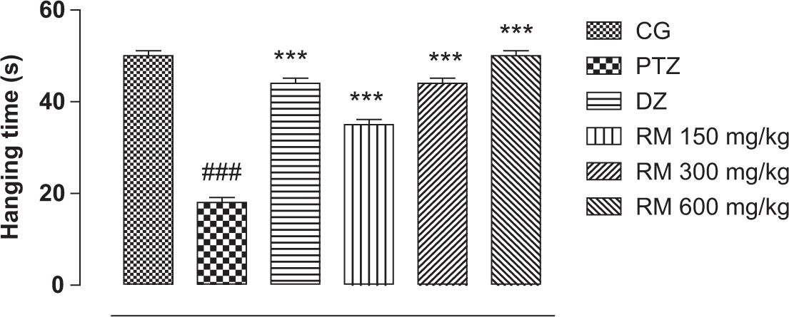
The wire hanging test results showed that compared to the standard group and animals in the RMLE 150, 300, and 600 mg/kg treatment groups and the control group, animals in the PTZ treatment group significantly (p < 0.05) fell from the wire hanging apparatus, demonstrating how RMLE therapy protected neuromuscular strength in a dose-dependent manner (Figure 2).
Figure 2. Effect of RMLE treatment on wire hanging test. Notes. The results (n = 6) are shown as mean ± SEM. The Boneferroni post hoc test with two-way ANOVA was used. ###p < 0.001, compared to the control group. **p < 0.01, compared to the disease control group, ***p < 0.001. CG: control group; PTZ: pentylenetetrazol, disease control; DZ: standard drug, diazepam; RM: R. moschata treatment groups.
Open field test
It was possible to study exploratory behavior, anxiety, and movement of the kindled epileptic animal model by providing open field environment. Compared to the control group, the kindled seizure model’s locomotion was reduced considerably (p < 0.05). RMLE therapy, however, significantly reduced the animals’ sad and aberrant neuropsychiatric behavior, which was not significantly different from the diazepam group. RMLE treatment groups underwent exploration while recovering in a dose-dependent manner (Figure 3).
Figure 3. Effect of RMLE treatment in open field test. Notes: The results (n = 6) are shown as mean ± SEM. The Boneferroni post hoc test with two-way ANOVA was used. ###p < 0.001, compared to the control group. **p < 0.01, compared to the disease control group, ***p < 0.001. CG: control group; PTZ: pentylenetetrazol, disease control; DZ: standard drug, diazepam; RM: R. moschata treatment groups.
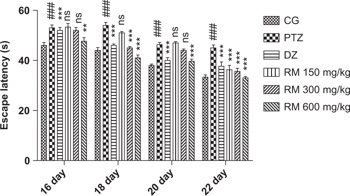
The MWM Test
Following the training session, measurements of animals’ escape latency were conducted on day 16, 18, 20, and 22. It was shown that kindled model mice took considerably (p < 0.05) more time to locate submerged platform and exit water. Compared to the standard group, treatment groups, and control groups, the escape latency of the intoxicated group on day 22 was noticeably longer with more time spent near the edge of the pool (Figure 4).
Figure 4. Effect of RMLE treatment on Morris’s water maze test. Notes: The results (n = 6) are shown as mean ± SEM. The Boneferroni post hoc test with two-way ANOVA was used. ###p < 0.001, compared to the control group. **p < 0.01, compared to the disease control group, ***p < 0.001. CG: control group; PTZ: pentylenetetrazol, disease control; DZ: standard drug diazepam; RM: R. moschata treatment groups.
Elevated plus Maze test
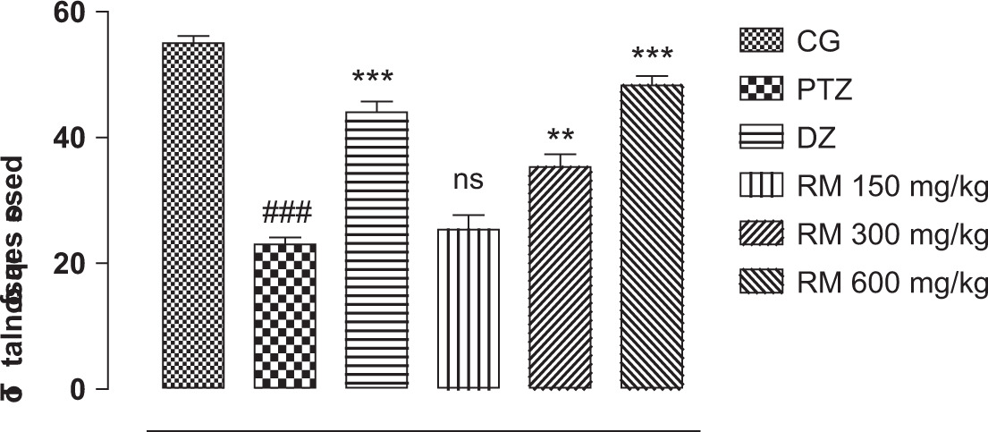
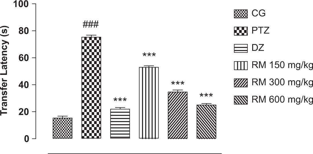
The kindled mice’s cognitive function was evaluated on day 22 of the experiment, and it was discovered that the transfer latency of PTZ-insulated mice was considerably (p < 0.05) stretched, compared to the groups that received the vehicle, and standard drug R. moschata 150, 300, and 600 mg/kg treatments. The performance of animals in terms of locomotion, exploration, and cognition was significantly improved by the RMLE therapy (Figure 5).
Figure 5. Effect of RMLE treatment on elevated plus maze test. Notes: The results (n = 6) are shown as mean ± SEM. The Boneferroni post hoc test with two-way ANOVA was used. ###p < 0.001, compared to the control group. **p < 0.01, compared to the disease control group, ***p < 0.001. CG: control group; PTZ: pentylenetetrazol, disease control; DZ: standard drug, diazepam; RM: R. moschata treatment groups.
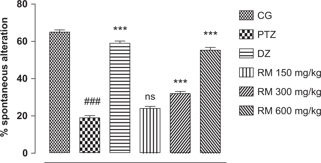
Y-Maze trial
To assess mice cognitive capabilities and propensity for exploration, the Y-maze experiment was used. We observed and kept track of the total the number of triads, number of arm entries, and the proportion of spontaneous modifications. Mice that received PTZ displayed a severe loss in cognition and mobility. PTZ-treated mice had considerably (p < 0.05) less arm entries, triads, and spontaneous alteration proportions, compared to the control, RMLE 150, 300, and 600 mg/kg treatment, and diazepam-treated mice groups. The current research showed that RMLE medication significantly reversed cognitive deterioration in a dose-dependent manner (Figure 6).
Figure 6. Effect of RMLE treatment on spontaneous alteration in Y-Maze test. Notes: The results (n = 6) are shown as mean ± SEM. The Boneferroni post hoc test with two-way ANOVA was used. ###p < 0.001, compared to the control group. **p < 0.01, compared to the disease control group, ***p < 0.001. CG: control group; PTZ: pentylenetetrazol, disease control; DZ: standard drug, diazepam; RM: R. moschata treatment groups.
Estimation of oxidative stress markers
Oxidative stress was brought on by PTZ therapy in the kindling mice model. The concentrations of the first-line antioxidant enzymes decreased considerably (p < 0.05 ). RMLE therapy (600, 300, and 150 mg/kg), however, (p < 0.05) restored considerably in a dose-dependent manner the levels of SOD, GSH, CAT, and proteins of treatment groups. In the kindling model, PTZ administration substantially (p < 0.05) elevated the levels of lipid peroxidation and nitrite. Treatment with RMLE reduced MDA and lipid peroxidation levels in a dose-dependent manner, although not significantly, compared to the control group (Table 1).
Table 1. Impact of RMLE therapy on nitrite levels, decreased glutathione protein, and oxidative stress indicators.
| Groups | Dose(mg/kg) | Superoxide dismutase(ug/mg of proteins) | Catalase(umol/mg) | Malonaldehyde(ug/mg) | Reduced glutathione(ug/mg of P) | Protein(ug/mg of P) | Nitrite(ug/mg of P) |
|---|---|---|---|---|---|---|---|
| CG | – | 4.32 ± 0.06 | 70.4 ± 0.20 | 3.79 ± 0.04 | 50.5 ± 0.23 | 29.30 ± 0.57 | 5.55 ± 0.02 |
| PTZ | 35 | 2.22 ± 0.55### | 48.5 ± 0.23### | 8.23 ± 0.05### | 40.1 ± 0.22### | 14.53 ± 0.23### | 7.75 ± 0.10### |
| DZ | 2 | 3.83 ± 0.05*** | 63.2 ± 0.08*** | 5.21 ± 0.05*** | 45.1 ± 0.32*** | 25.21 ± 0.08*** | 4.69 ± 0.03*** |
| Treatment(RMLE) | 150 | 3.87 ± 0.06*** | 59.8 ± 0.04*** | 4.26 ± 0.04*** | 43.5 ± 0.02*** | 22.23 ± 0.09*** | 5.79 ± 0.07*** |
| 300 | 4.19 ± 0.05*** | 67.8 ± 0.05*** | 3.81 ± 0.06*** | 46.3 ± 0.06*** | 26.8 5 ± 0.05*** | 4.66 ± 0.07*** | |
| 600 | 5.09 ± 0.04*** | 69.6 ± 0.27*** | 3.60 ± 0.27*** | 48.1 ± 0.06*** | 27.17 ± 0.02*** | 3.88 ± 0.06*** |
Notes: The results (n = 6) are shown as mean ± SEM. The Boneferroni post hoc test with two-way ANOVA was used. ###p < 0.001 compared to the control group. ***p < 0.001 compared to the disease control group.
CG: normal Control group; PTZ: pentylenetetrazol, disease control; DZ: standard drug diazepam group.
Estimation of catalase level
In estimation of catalase level, considerable (p < 0.001) decrease was found in the disease control group, compared to the control group. The diazepam group meaningfully (p < 0.01) overturned alteration in catalase level. When RMLE extract was administered for consecutive 22 days, momentous (p < 0.001) dose-dependent increase was observed in catalase level at RMLE 150, 300 and 600 mg/kg (Table 1).
Super oxide dismutase (SOD) level
In comparison to the control group, a decrease in the SOD level of brain tissue homogenate was observed in the disease control group (p < 0.001). The diazepam-treated group expressively (p < 0.01) reversed change in SOD level induced by PTZ. Significant (p < 0.001) improvement was detected at 150 mg/kg. In addition, extract dose at 300 mg/kg (p < 0.01) and 600 mg/kg (p < 0.001) also showed substantial improvement (Table 1).
Estimation of reduced glutathione level
Pentylenetetrazol caused momentous (p < 0.001) lessening of the GSH level in brain homogenate of disease animals, compared to the control group. GSH level meaningfully (p < 0.001) reached near to control value in standard group. The GSH level was significantly recovered in treatment groups (p < 0.001) at all dose levels shown in Table 1.
Evaluation of glutathione peroxidase level
In disease animals, a noteworthy (p < 0.001) lessening of GPx level in brain tissue homogenate was observed, compared to the control group. GPx level meaningfully (p < 0.001) reached near to the control value in standard group. Substantial (p < 0.001) improvement was detected at RMLE dose of 150 mg/kg. Correspondingly, extract dose of 300 mg/kg (p < 0.01) and 600 mg/kg (p < 0.001) also presented substantial improvement (Table 1).
Determination of malonaldehyde level
Noteworthy (p < 0.001) decline was observed in the MDA level of brain tissue homogenate of the disease-induced group, compared to the control group. MDA concentration expressively (p < 0.001) reached near the control value in the standard group. Significant (p < 0.001) improvement was detected at RMLE dose of 150 mg/kg. Extract dose at 300 mg/kg (p < 0.01) and 600 mg/kg (p < 0.001) also displayed substantial improvement when RMLE extract was administered for consecutive 22 days (Table 1).
Estimation of total protein
Protein level decreased considerably (p < 0.001) in disease group animals. Standard control group significantly (p < 0.001) increased protein level. Significant improvement in total protein level was detected in the RMLE treatment groups of 150 mg/kg (p < 0.05), 300 mg/kg (p < 0.01), and 600 mg/kg (p < 0.001) (Table 1).
Quantification of neurotransmitters level in brain
Quantification of acetyl cholinesterase
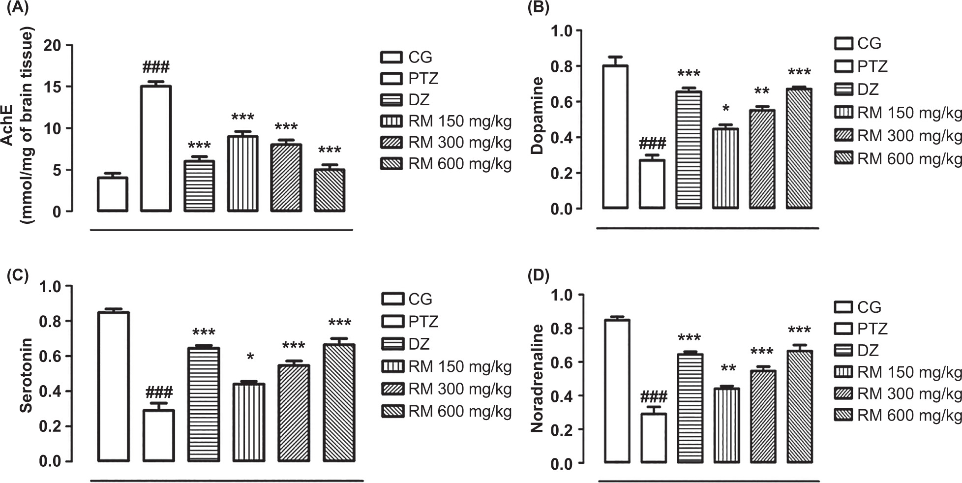
Owing to a considerable increase in the amount of acetyl cholinesterase (p < 0.05), the level of acetylcholine (ACh) was noticeably lowered in PTZ-affected mice. RMLE therapy, however, dose-dependently restored the level of ACh and cognitive function via modulating the level of acetyl cholinesterase in the treatment group that was nonsignificant in contrast to the disease control and control groups (Figure 7A).
Figure 7. Effect of RMLE treatment on (A) acetylcholinesterase activity and levels of (B) dopamine, (C) serotonin, and (D) noradrenaline. Notes: The results (n = 6) are shown as mean ± SEM. The Boneferroni post hoc test with two-way ANOVA was used. ###p < 0.001, compared to the control group. **p < 0.01, compared to the illness control group, ***p < 0.001. CG: control group; PTZ: pentylenetetrazol, disease control; DZ: standard drug diazepam; RM: R. moschata treatment groups.
Estimation of dopamine level in brain
Dopamine level measured in brain tissue homogenate decreased substantially (p < 0.001) in disease control animals. Dopamine level was restored significantly (p < 0.001) in standard and 600 mg/kg dose group. RMLE groups presented a substantial dose-dependent recovery of dopamine level (p < 0.05) at 150 mg/kg and (p < 0.001) at 300 mg/kg, compared to the disease control group (Figure 7B).
Estimation of serotonin level
Decrease in serotonin level in epilepsy is correlated to mood disturbances and dyskinesia. Serotonin level pointedly (p < 0.001) reduced in PTZ-treated group, rather being normal in the control group. RMLE extract-treated groups at 150 mg/kg, 300 mg/kg, and 600 mg/kg showed significant improvement in serotonin levels. Standard group also showed remarkable (p<0.001) increase in serotonin level (Figure 7C).
Estimation of noradrenaline level
The noradrenaline level estimated in brain tissue homogenate was considerably (p < 0.001) reduced in PTZ-treated groups, compared to the control group animals. Noradrenalin level was restored pointedly (p < 0.001) in the standard group. Animals treated with RMLE extract 150 mg/kg, 300 mg/kg, and 600 mg/kg exhibited a dose-dependent recovery of noradrenaline (p < 0.05), compared to the disease control group (Figure 7D).
Hematological examination
The control group values of hemoglobin (Hb, 13.5±0.51), hematocrit (HCT, 7.82±0.02), and red blood cells (RBC, 47.4±0.57) remained within the normal range, compared to RMLE treatment animals (Hb, M: 12–17: F: 12–16, and HCT 35–55%). However, Hb (11.2±0.57), RBC (4.6±0.12), and hematocrit (42.3±0.57) dropped markedly (p < 0.001) in the PTZ treatment group. White blood cells (WBC, 13±0.57), neutrophils (77±0.52), lymphocytes (23.5±0.53), monocytes (05±0.32), and platelets (1,147±8.80) increased significantly (p < 0.001) in PTZ treatment group, compared to the control group values of WBC (10.6±0.32), neutrophils (69.5 ± 0.57), lymphocytes (18.3 ± 0.57), monocytes (03 ± 0.39), and platelets (344 ± 8.27), but were in normal range (Table 2). At various doses, RMLE extract had no negative impact on hematological markers.
Table 2. Impact of RMLE extract on red blood cells and white blood cells.Notes. The results (n = 6) are shown as mean ± SEM. The Boneferroni post hoc test with two-way ANOVA was used. ###p < 0.001, compared to the control group. **p < 0.01, compared to the illness control group, ***p < 0.001.
| Parameters | Units | CG | PTZ | DZ | Treatment (RMLE) | ||
|---|---|---|---|---|---|---|---|
| 150 mg/kg | 300 mg/kg | 600 mg/kg | |||||
| Hb | g/dL | 13.5 ± 0.51 | 11.2 ± 0.57### | 14.5 ± 0.57*** | 12.5 ± 0.52*** | 14.1 ± 0.01** | 11.2 ± 0.53*** |
| RBC | ×10-6/uL | 7.82 ± 0.02 | 4.6 ± 0.12### | 10.55 ± 0.05** | 7.81 ± 0.01* | 7.56 ± 0.05** | 8.4 ± 0.12*** |
| Hematocrit | % | 47.4 ± 0.57 | 42.3 ± 0.57### | 58.7 ± 0.57*** | 47.3 ± 0.57*** | 45.2 ± 0.87*** | 45.5 ± 0.88** |
| MCV | % | 60.6 ± 0.53 | 85.1 ± 0.31### | 55.6 ± 0.57*** | 64.4 ± 0.57*** | 59.8 ± 0.57** | 71.6 ± 0.33*** |
| MCH | Pg | 17.3 ± 0.57 | 21.5 ± 0.88### | 15.6 ± 0.57*** | 36.3 ± 0.57*** | 18.7 ± 0.57* | 26.4 ± 0.57*** |
| MCHC | % | 25.5 ± 0.57 | 28.2 ± 0.57### | 28.1 ± 0.57*** | 28.5 ± 0.57*** | 28.0 ± 0.57* | 29.0 ± 0.57** |
| RDW-CV | % | 16.7 ± 0.33 | 17.2 ± 0.57### | 21.2 ± 0.31* | 20.7 ± 0.33*** | 12.5 ± 0.57*** | 23.2 ± 0.57*** |
| WBC | ×10-3/µL | 10.6 ± 0.32 | 13 ± 0.57### | 5.8 ± 0.33** | 4.0 ± 0.57* | 7.0 ± 0.57*** | 9.7 ± 0.32*** |
| Neutrophils | % | 69.5 ± 0.57 | 77 ± 0.52### | 68.5 ± 0.57*** | 70.5 ± 0.57** | 60.5 ± 0.57** | 71.4 ± 0.57*** |
| Lymphocytes | % | 18.3 ± 0.57 | 23.5 ± 0.53### | 20.5 ± 0.51*** | 22.5 ± 0.54* | 32.5 ± 0.57*** | 22.6 ± 0.51*** |
| Monocytes | % | 03 ± 0.39 | 05 ± 0.32### | 07 ± 0.32** | 02 ± 0.33*** | 04 ± 0.34** | 04 ± 0.32*** |
| Eosinophils | % | 04 ± 0.32 | 03 ± 0.42### | 04 ± 0.32* | 05 ± 0.34*** | 03 ± 0.32** | 02 ± 0.33*** |
| Platelet count | ×10-3/µL | 344 ± 8.27 | 1147 ± 8.80### | 978 ± 13.2* | 832 ± 4.75* | 1075 ± 8.21** | 965 ± 13.4*** |
CG: control group; PTZ: pentylenetetrazol disease control; DZ: standard drug diazepam; RM: R. moschata treatment groups.
Effect of RMLE extract on renal function test (RFT)
Compared to the values of the control group (22±0.32), blood urea level (40±0.57) in the PTZ treatment group was increased significantly (p < 0.001). RMLE extract-treated groups at the doses of 150 mg/kg, 300 mg/kg, and 600 mg/kg showed notable (p < 0.001) respective decrease in blood urea level of 37±0.33, 29 ±0.57, and 24±0.53 (Table 3).
Table 3. Effect of RMLE extract on renal function test (RFT) and liver function test (LFT).
| Groups | Dose(mg/kg) | Blood urea (mg/dL) | Serum creatinine(md/dL) | Serum bilirubin(mg/dL) | ALT | AST | ALP |
|---|---|---|---|---|---|---|---|
| CG | - | 22 ± 0.32 | 0.8 ± 0.03 | 0.9 ± 0.06 | 21 ± 0.06 | 28 ± 0.01 | 115 ± 0.02 |
| PTZ | 1 | 40 ± 0.57### | 1.2 ± 0.02### | 0.23 ± 0.01### | 32 ± 0.13### | 37 ± 0.02### | 210 ± 0.03### |
| DZ | 100 + 25 | 18 ± 0.32*** | 0.7 ± 0.01*** | 0.7 ± 0.02** | 19 ± 0.03** | 24 ± 0.01*** | 208 ± 0.04** |
| Treatment(RMLE) | 150 | 37 ± 0.33** | 1.1 ± 0.02* | 0.20 ± 0.04*** | 27 ± 0.05*** | 35 ± 0.01*** | 194 ± 0.01** |
| 300 | 29 ± 0.57*** | 0.9 ± 0.01** | 0.13 ± 0.02* | 25 ± 0.01** | 32 ± 0.01*** | 188 ± 0.03*** | |
| 600 | 24 ± 0.53*** | 0.6 ± 0.03*** | 0.10 ± 0.01*** | 24 ± 0.06*** | 30 ± 0.001*** | 157 ± 0.02*** |
Notes: The results (n = 6) are shown as mean ± SEM. The Boneferroni post hoc test with two-way ANOVA was used. ###p < 0.001, compared to the control group. **p < 0.01, compared to the illness control group, ***p < 0.001. CG: control group; PTZ: pentylenetetrazol, disease control; DZ: standard drug diazepam; RM: R. moschata treatment groups.
The serum creatinine level (1.2±0.02) in PTZ treatment groups was noticeably (p < 0.001) higher than the control group value (0.8±0.03). Treatment with RMLE at the doses of 150 mg/kg, 300 mg/kg, and 600 mg/kg resulted in significant dose-dependent (p < 0.001) lowering of the respective serum creatinine level of 1.1±0.02, 0.9±0.02, and 0.6±0.03 (Table 3).
Effect of RMLE extract on liver function test (LFT)
The PTZ treatment group had a substantially higher serum bilirubin level (0.23±0.01) compared to the control group (0.9±0.06) (p < 0.001).
RMLE extract treatment at the doses of 150 mg/kg, 300 mg/kg, and 600 mg/kg resulted in a dose-dependent significant (p < 0.001) decrease in respective blood bilirubin levels of 0.20±0.04, 0.13±0.02, and 0.10±0.01 (Table 3).
In PTZ treatment group, alanine transferase (ALT) level (32 U/L) increased considerably (p < 0.001), compared to the control group (21 U/L). Alanine transferase level decreased considerably (p < 0.001) at RMLE extract doses of 150 mg/kg, 300 mg/kg, and 600 mg/kg (Table 3).
Histopathological analysis
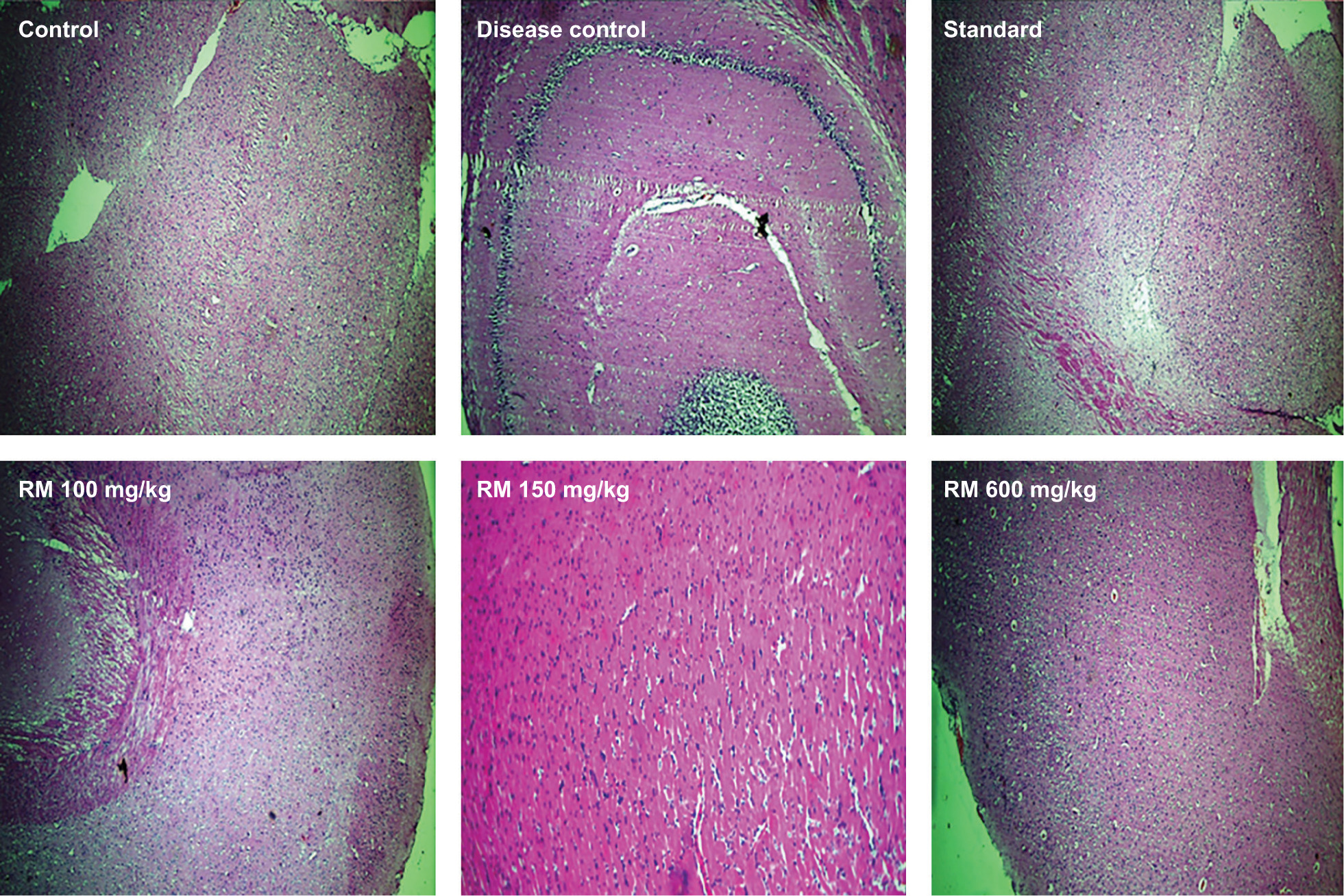
The analyzed control groups showed no granules according to histopathological analysis. However, hippocampus slices of the PTZ-treated group showed aberrant neuronal organization, neuronal loss and degeneration, presence of apoptotic bodies, proliferation of inflammatory mediators, and mild edema. However, RMLE treatment at doses of 150 mg/kg, 300 mg/kg, and 600 mg/kg significantly increased the structural integrity of the hippocampus (Figure 8).
Figure 8. Effect of RMLE on the brain histopathology of various experimental groups.
Histopathology of heart tissues
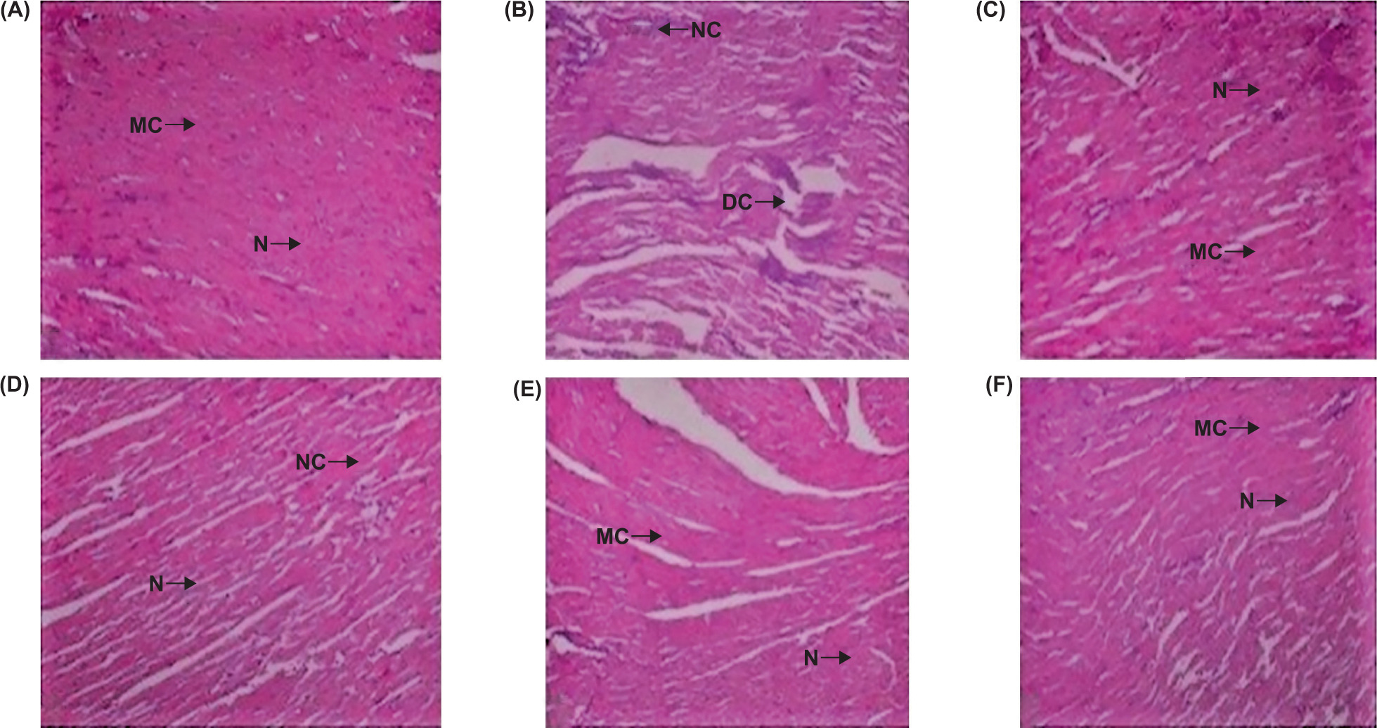
Treatment with RMLE had no discernible negative effect on the histopathology of vital organs. The cardiac fiber architecture of the extract treated groups was similar to that of the control group. However, in the PTZ treatment group, vascular congestion in myocardial fibers was moderately severe. The histopathology of cardiac muscles demonstrated that RMLE had no adverse effect (Figure 9).
Figure 9. Impact of RMLE therapy on the histopathology of cardiac tissues. (A): normal control group; (B): PTZ disease control; (C): DZ: diazepam standard drug; (D–F): respective RMLE treatment group of 150 mg/kg, 300 mg/kg, and 600 mg/kg. CN: central nuclei; MF: myocardial fibrils.
Histopathology of kidney tissues
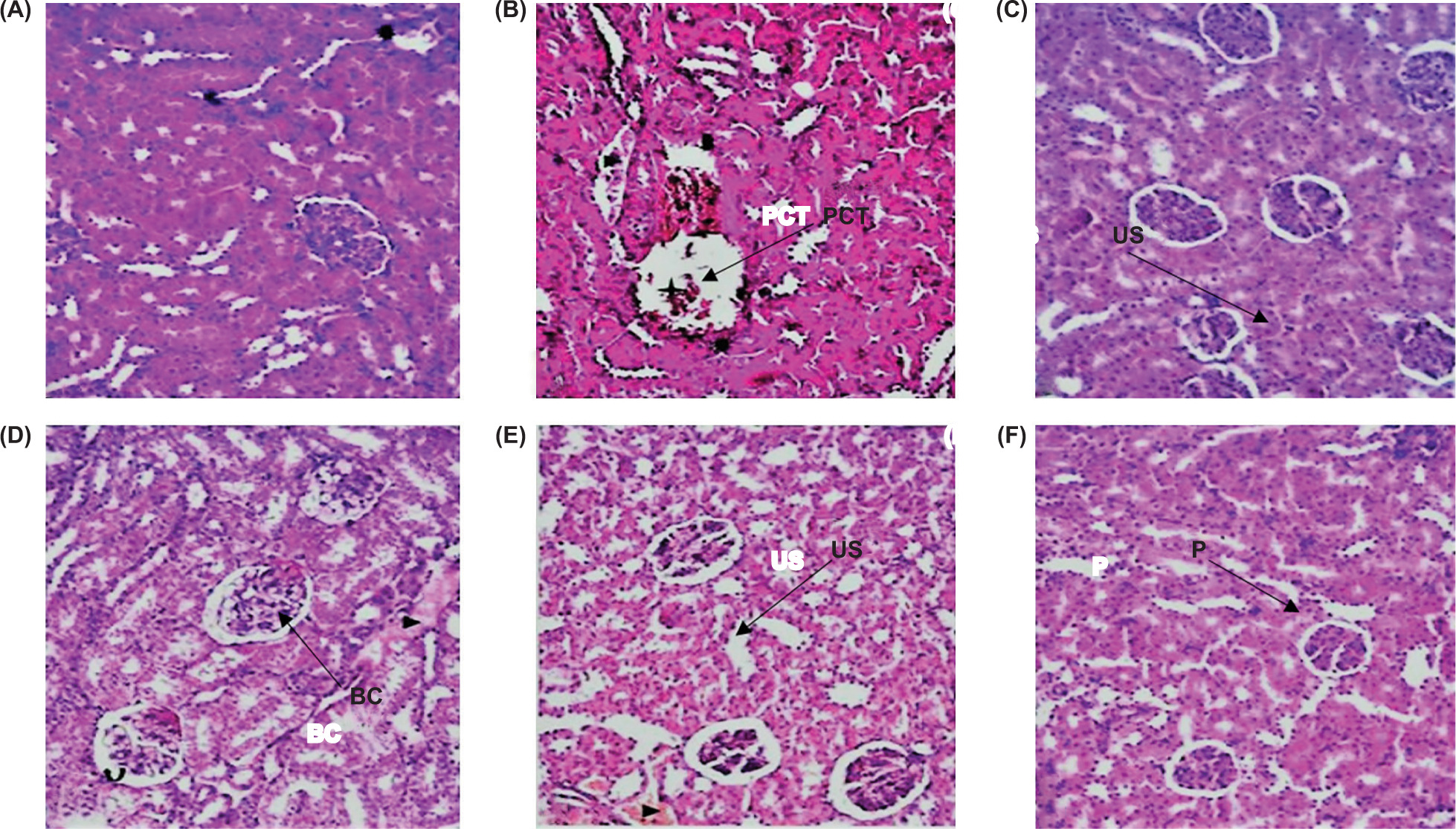
RMLE extract had no toxic effects on the histology of nephrons. The therapy groups exhibited normal renal parenchyma, much like the control group. However, renal parenchyma and kidney cell nuclei in the PTZ treatment group presented moderate levels of necrotic alterations and congestion (Figure 10).
Figure 10. Effect of RMLE on the histopathology of mice kidney tissues. (A) normal control group; (B) PTZ disease control; (C) DZ: diazepam standard drug; (D–F) respective RMLE treatment groups of 150 mg/kg, 300 mg/kg, and 600 mg/kg. US: urinary space; BC: Bowman’s capsule; P: podocytes; PCT: proximal convoluted tubules.
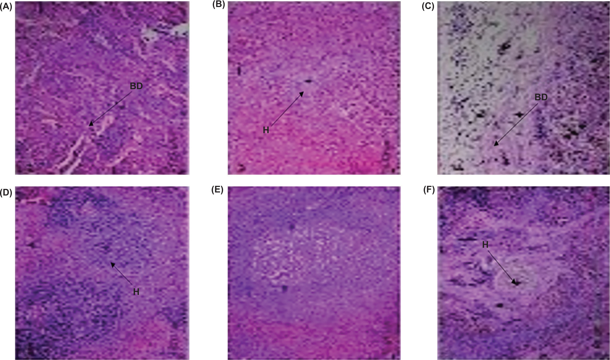
Histopathology of liver tissues
The liver in the control group was compact, contained hepatocytes that were typical in shape and connected to the portal vein, and was free of fibrosis. The parenchyma cells in the livers of the PTZ treatment group showed minor vascular congestion and vacuolization. Hepatic vacuolization was prevalent in parenchyma cells of the control group. RMLE at a dosage of 600 mg/kg revealed normal epithelial cells and mildly congested hepatocytes (Figure 11).
Figure 11. Effect of RMLE on the histopathology of liver tissues. (A) normal control group; (B) PTZ disease control; (C) DZ: diazepam, standard drug; (D–F): respective RMLE treatment groups of 150 mg/kg, 300 mg/kg, and 600 mg/kg.
Histopathology of lungs tissues
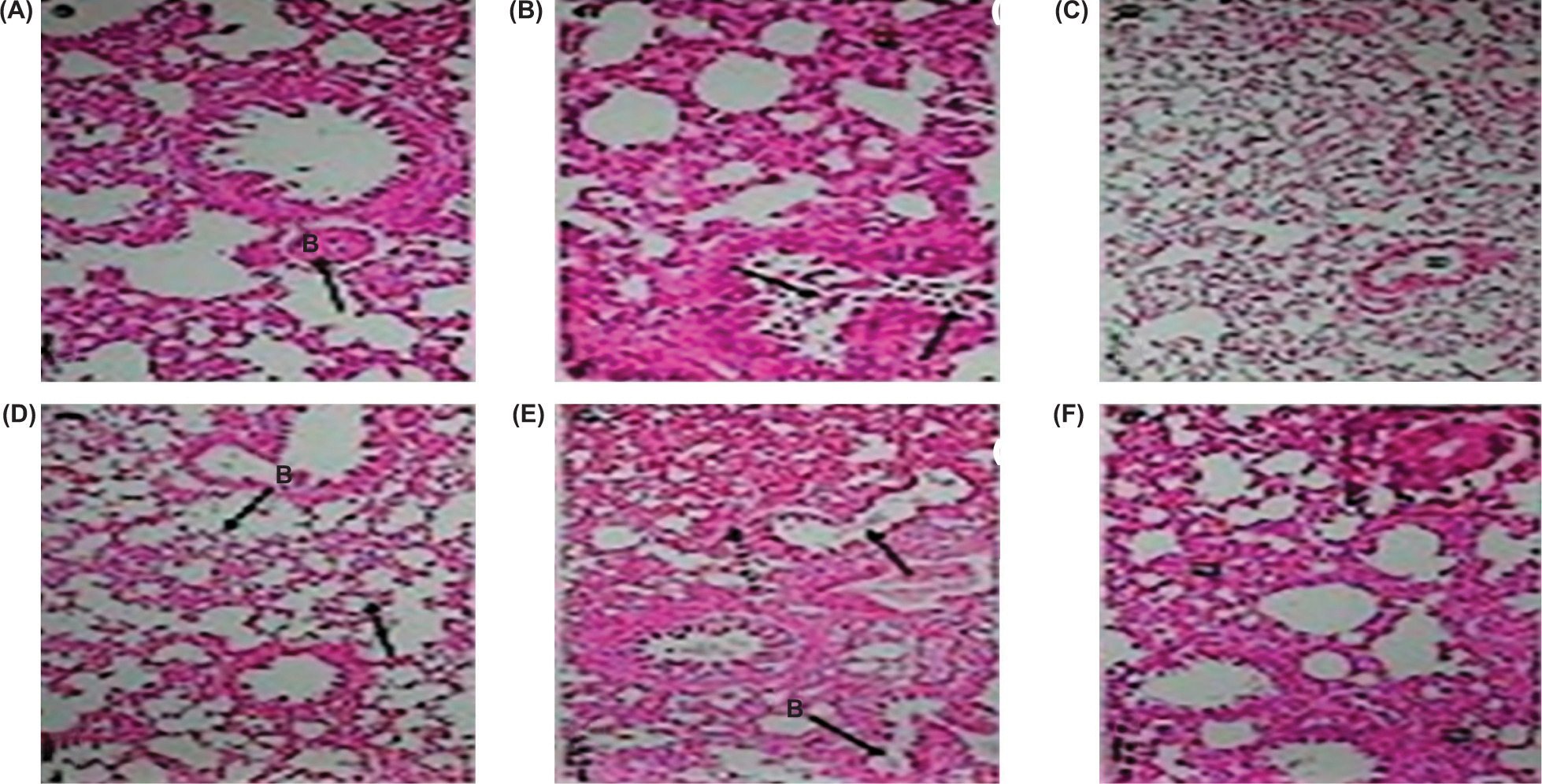
The lungs’ alveoli, bronchi, blood vessels, and epithelium of the control group were in normal shape. The PTZ treatment group showed lung congestion. The bronchi and alveoli in the control group were normal. No histopathologically damaging symptoms were observed in the lungs tissues of the RMLE-treated groups (Figure 12).
Figure 12. Effect of RMLE on the histopathology of lung tissues. (A) normal control group; (B) PTZ disease control; (C) DZ: diazepam, standard drug; (D–F) respective RMLE treatment groups of 150 mg/kg, 300 mg/kg, and 600 mg/kg.
Discussion
The associated diseases, psychosis, learning and memory impairment, and cognitive decline coexist with anticonvulsant effects (Upaganlawar et al., 2021). Seizures are linked to mood disorders, such as sadness, anxiety, and psychosis. The precise neurobiological processes underpinning the link between epilepsy and aberrant behavior are not understood completely (Kanner, 2007).
Given the widespread occurrence of epilepsy with anticonvulsant effects and the declining effectiveness of currently available medications, it is crucial to explore new herbal treatments that may offer significant efficacy and comparatively reduced adverse effects. Recently, treatment with antioxidants, specifically herbal treatment, has been studied widely to prevent the occurrence of different diseases and adverse effects (Sureda et al., 2023). Therefore, in a PTZ kindling model of epilepsy in mice, we investigated the therapeutic effects of R. moschata’s aqueous ethanolic extract to control seizures, address cognitive impairments, and reduce oxidative stress.
Following PTZ injection, kindled mice underwent behavioral phenotyping utilizing behavioral test batteries. Locomotor activity, learning and cognitive performance, emotions, dejection, and anxiousness like psychopathology, a series of behavioral batteries were recorded during the study (Kanner, 2022). The mice employed in the chemically induced kindling paradigm demonstrated reduced locomotor movement, as demonstrated by the raised plus maze activity, MWM test, and open field test. However, as observed in the wire hanging challenge, the neuromuscular strength was also noticeably reduced in kindled mice (Anisman and McIntyre, 2002). The results of behavioral investigation showed a high correlation between the onset of seizures and neurodegenerative changes that impaired learning ability and cognitive function (Meng et al., 2014). RMLE therapy reversed behavioral impairments, decreased locomotor function and activity, learning, and memory deterioration in treatment groups, compared to the PTZ-induced mice (Ahmadian et al., 2019). This is likely because of its neuroprotective and antioxidant capabilities similar to Meng et al.’s (2014) experimental study, where 150 mg/kg of resveratrol significantly enhanced behavioral changes, neuronal count, and cognitive function in the MWM challenge (Hannesson et al., 2004). Animals treated with all three doses of RMLE also recovered from the behavioral and biochemical abnormalities brought on by PTZ administration.
Phytochemical characterization through HPLC analysis in our earlier studies demonstrated the presence of salicylic acid, gallic acid, p-coumaric acid, and vanillic acid (Agarzae et al., 2025; Alotaibi et al., 2025). Among these compounds, gallic acid has proven neuroprotective role through antioxidant and anti-inflammatory mechanisms (Yazğan, 2024) and vanillic acid also showed anticonvulsant potential by reduction of PTZ-induced seizures in mice because of impairment in neurotransmission (Farbood et al., 2023). p-Coumaric acid, a natural polyphenolic found in several plants, exhibits neuroprotective effects by combating oxidative stress and inflammation within the brain (Sakamula and Thong-Asa, 2018). It has been demonstrated that salicylic acid, a well-known neuroprotectant and strong anti-inflammatory medication, guards against toxicity brought on by other dopaminergic neurotoxins (Thrash-Williams et al., 2016).
Rosa moschata leaf extract dramatically reduced neuroinflammation biomarkers, aberrant behavior, and improved cognitive function, similar to that of arbutin (Shekh-Ahmad et al., 2019). According to reports, the redox potential is altered in status epilepticus, causing a decrease in adenosine triphosphate (ATP) synthesis and a collapse of brain bioenergetics. Chronic convulsions or seizures cause oxidative stress, which damages proteins, lipids, DNA, and causes mitochondrial malfunctioning (Lin et al., 2020). An essential technique for a thorough and improved understanding of seizure pathophysiology and development is the induction of seizures in animal models. PTZ oral administration induced mitochondrial malfunctioning, protein oxidation, neuronal death, lipid peroxidation, and oxidative stress (Shakeel et al., 2017). PTZ produces reactive oxygen species (ROS) and lipid peroxidation in the hippocampus and frontal cortex as a result of its excitotoxic activation. The blocking of nitric oxide synthase (NOS) activity also could cause vasoconstriction with formation of ROS (Selamoglu-Talas et al., 2013). Oxidative stress caused by imbalance of free radicals leads to many chronic brain diseases (Shah et al., 2017). Free radical production increased due to ongoing seizures, additional oxidative injury, and neuronal deterioration (Samokhina and Samokhin, 2018). In the current study, sub-convulsive PTZ doses were used to maintain oxidative stress (Nascimento et al., 2005). Antioxidant enzymes catalase, SOD, GSH reductase, and GPx are normalized as a result of the unbalanced oxidant/antioxidant equilibrium (Diniz et al., 2015).
One prevalent mechanism of cellular damage in a variety of acute neurological events, such as seizures and illnesses like Alzheimer’s disease, is oxidative stress in the brain.
Particularly susceptible to lipid peroxidation are membrane lipids, which are abundant in unsaturated fatty acids, such as arachidonic acid. This process damages the membrane and reduces its functionality. Normally, SOD partially mitigate the harmful effects of oxidative stress and free radicals (Poon et al., 2004).
In patients having convulsions, serum levels of antioxidants are reduced, thus leading to increased lipid peroxidation because of elevated levels of free radicals. PTZ causes seizures by inducing oxidative stress, and administration of antioxidants significantly reduced both oxidative stress and PTZ-induced seizures. According to other sources, PTZ may cause the membrane structure to lyse, which releases free radicals and lipid peroxidases.
The oxidative stress effects of PTZ induction were highlighted in our study by the PTZ-treated group’s significantly elevated MDA levels, an indication of lipid peroxidation, and lower levels of antioxidant enzyme SOD. The first-line antioxidant enzyme level dramatically increased after RMLE therapy, restoring the seizure threshold.
The levels of brain neurotransmitters, such as adrenaline, dopamine, and serotonin, were also disrupted due to induction of seizures. RMLE therapy, on the other hand, controlled brain neurotransmitter levels and restored seizure latency in the treated mice. Oxidative stress was increased by status epilepticus, which led to neuronal death and neurodegeneration (Muscatello et al., 2018). Increased concentrations of neuro-inflammatory biomarkers are connected with the neurodegeneration brought on by epileptic convulsions. However, the elevated expression is apparent in treated animals because of RMLE protective functioning. Acetyl cholinesterase enzyme catalyzes ACh hydrolysis. It restores cholinergic nerve pathways by having a role in neurotransmission. It is essential for determining the neurotoxic effects of xenobiotics that cause the central and peripheral nervous systems to degenerate (Waiskopf and Soreq, 2015). In particular, because of compromised nerve conduction, acetylcholinesterase (AChE) inhibition, and the buildup of ACh in synaptic route can result in life-threatening symptoms, such epileptic convulsions, coma, and death (Balali-Mood and Saber, 2012). In the current study, AChE levels were found to be elevated in the brain tissues of PTZ-induced illness group. The RMLE-treated groups’ AChE levels were considerably lower than those of the PTZ and normal control groups. The levels of AChE in serum and brain tissue were comparable.
The expression of neurodegenerative proteins is strongly correlated with seizure activity.
Previous published findings clearly demonstrated a relationship between neurodegeneration and epilepsy and overexpression of α-synuclein. The increased mRNA expression of neurodegenerative proteins was reported, which was consistent with earlier results in the PTZ-induced kindling paradigm (Paudel et al., 2020). RMLE therapy reduced neurodegenerative pathogenesis, neuronal death, and neuroinflammation in a dose-dependent manner (Choi et al., 2020).
Conclusion
The present study provides strong experimental evidence that RMLE demonstrates significant anticonvulsant, neuroprotective, and antioxidative properties in a PTZ-kindled mice model of epilepsy. Administration of RMLE significantly reduces seizure severity, improved cognitive and behavioral performance, restored neurotransmitter balance, and enhanced endogenous antioxidant enzyme activity while reducing oxidative stress markers. Histopathological analyses confirm the neuroprotective potential of RMLE extract, showing preserved neuronal integrity and minimal tissue damage in vital organs. Additionally, RMLE demonstrates a favorable safety profile, with no significant adverse effects on hematological, renal, or hepatic parameters. Collectively, these findings validate the traditional use of Rosa moschata in neurological disorders and highlight its potential as a promising multi-target therapeutic candidate for epilepsy management and associated comorbidities. Further studies, including isolation of active constituents and clinical trials, are required to elucidate its mechanism of action and therapeutic applicability in humans.
Acknowledgment
The authors thank the Ongoing Research Funding Program (ORF-2025-1210), King Saud University, Riyadh, Saudi Arabia, for financial support. The authors also acknowledge Universitas Diponegoro, Indonesia, through its Adjunct Professor, World-Class University Program (2025), and Syarif Hidayatullah State Islamic University Jakarta, Indonesia, through its International Research Fellowship Program (2025).
Author Contributions
US, MAS, ASA and NKA conceived and supervised the study. HA, HG, MF, AMA, AA and WSA performed the experiments. IA, NAA, AKK, AGA, AA, NA, ALNA and PA analyzed and interpreted the results. All authors have equally contributed to writing, editing, and revising the final draft.
Conflicts of Interest
None.
Funding
This research was funded by Ongoing Research Funding program, (ORF-2025-1210), King Saud University, Riyadh, Saudi Arabia.
REFERENCES
Abbas, A., Mubeen, M., Zheng, H., Sohail, M.A., Shakeel, Q., Solanki, M.K., . . . Hussain, S. 2022. Trichoderma spp. genes involved in the biocontrol activity against Rhizoctonia solani. Front Microbiol. 13:884469. 10.3389/fmicb.2022.884469
Agarzae, N.K., Saleem, U., Alsharif, I., Safdar, R., Chaudhary, Z., Alqahtani, M.J., . . Silva, S. 2025. Rosa moschata leaf extract ameliorates Alzheimer’s disease in AlCl3 + D-galactose induced Alzheimer’s rat model via modulation of neuroinflammatory biomarkers and suppression of oxidative stress markers. Ital J Food Sci. In Press. 10.15586/ijfs.v37i4.3123
Ahandani, E.A., Ozdemir, B., Hajipour, S., Selamoglu, Z., Riaz, A., Issa, H. Y., ... Madhumitha Sri, R.M. 2023. Use of natural products in preventive medicine and healthy life. J Prev Med Holistic Health. 9(1):5–9. 10.18231/j.jpmhh.2023.003
Ahmadian, S.R., Ghasemi-Kasman, M., Pouramir, M., and Sadeghi, F.J.N. 2019. Arbutin attenuates cognitive impairment and inflammatory response in pentylenetetrazol-induced kindling model of epilepsy. Neuropharmacology. 146:117–127. 10.1016/j.neuropharm.2018.11.038
Akyüz, E., Köklü, B., Ozenen, C., Arulsamy, A., and Shaikh, M.F.J.C. 2021. Elucidating the potential side effects of current anti-seizure drugs for epilepsy. 19(11):1865. 10.2174/1570159X19666210826125341
Alotaibi, B.S., Saleem, U., Farrukh, M., Chaudhary, Z., Anwar, N., Alsharif, I., . . . Al-Qahtani, W.S. 2025. Rosa moschata ameliorates haloperidol-induced Parkinson’s disease via reduction of neurodegeneration and oxidative stress. Asian Pacific J Trop Biomed. 15(1):24–33. 10.4103/apjtb.apjtb_451_24
Anisman, H., and McIntyre, D.C. 2002. Conceptual, spatial, and cue learning in the Morris water maze in fast or slow kindling rats: attention deficit comorbidity. 22(17):7809–7817. 10.2174/1570159X19666210826125341
Balali-Mood, M., and Saber, H. 2012. Recent advances in the treatment of organophosphorous poisonings. Iran J Med Sci. 37(2):74.
Benbadis, S.J.E. 2009. The differential diagnosis of epilepsy: a critical review. 15(1):15–21. 10.1016/j.yebeh.2009.02.024
Choi, J., Kim, S.Y., Kim, H., Lim, B.C., Hwang, H., Chae, J.H., . . . Shin, J.-S. 2020. Serum α-synuclein and IL-1β are increased and correlated with measures of disease severity in children with epilepsy: potential prognostic biomarkers? 20(1):1–11. Retrieved from https://link.springer.com/article/10.1186/s12883-020-01662-y
Diniz, T.C., Silva, J.C., Lima-Saraiva, S.R.G.d., Ribeiro, F.P.R.d.A., Pacheco, A.G.M., de Freitas, R.M., . . . Longevity, C. 2015. The role of flavonoids on oxidative stress in epilepsy. 10.1155/2015/171756
Farbood, Y., Sedeh, S.S., Sarkaki, A., Ahangarpour, A., and Dianat, M. 2023. Protective effect of vanillic acid against pentylenetetrazol-induced convulsions in male rats. Jundishapur J Physiol. 2(2):99–108. 10.3295/JJP.2023.2.2.99
Fischer, R., Maier, O.J.O.m., and Longevity, C. 2015. Interrelation of oxidative stress and inflammation in neurodegenerative disease: role of TNF. 10.1155/2015/610813
Getnet, A., Woldeyohannes, S.M., Bekana, L., Mekonen, T., Fekadu, W., Menberu, M., . . . Belete, H.J.B. 2016. Antiepileptic drug nonadherence and its predictors among people with epilepsy. 10.1155/2016/3189108
Gharaghani, M., Jafarian, H., Hatami, M., Shabanzadeh, M., and Mahmoudabadi, A.Z. 2022. Evaluation of catalase activity of clinical and environmental isolates of Aspergillus species. Iran J Microbiol. 14(1):133. 10.18502/ijm.v14i1.8815
Hannesson, D., Wallace, A., Pollock, M., Corley, S., Mohapel, P., and Corcoran, M.J.E.r. 2004. The relation between extent of dorsal hippocampal kindling and delayed-match-to-place performance in the Morris water maze. Epilepsy Res. 58(2–3):145–154. 10.1016/j.eplepsyres.2004.01.004
Hesdorffer, D.C., Benn, E.K., Cascino, G.D., and Hauser, W.A.J.E. 2009. Is a first acute symptomatic seizure epilepsy? Mortality and risk for recurrent seizure. 50(5):1102–1108. 10.1111/j.1528-1167.2008.01945.x
Hooijmans, C.R., Buijs, M., Struijs, F., Som, T., Karim, N., Scheffer, G.-J., and Malagon, I.J.S.R. 2023. Exposure to halogenated ethers causes neurodegeneration and behavioural changes in young healthy experimental animals: a systematic review and meta analyses. Sci Rep, 13(1):8063. 10.1038/s41598-023-35052-4
Hughes, J., Devinsky, O., Feldmann, E., and Bromfield, E.J.S. 1993. Premonitory symptoms in epilepsy. 2(3):201–203. 10.1016/S1059-1311(05)80128-1
Ju, J.-H., Yoon, Y.-H., Shin, S.-H., Ju, S.-Y., and Yeum, K.-J. J. H. 2022. Recent trends in urban agriculture to improve bioactive content of plant foods. 8(9):767. 10.3390/horticulturae8090767
Kandeda, A.K., Moto, F.C.O., Ayissi, R.E.M., Omam, J.P.O., Ojong, L., and Bum, E.N. 2021. Pergularia daemia hydro-ethanolic extract protects against pentylenetetrazol kindling-induced seizures, oxidative stress, and neuroinflammation in mice. J Ethnopharmacol. 279:114338. 10.1016/j.jep.2021.114338
Kanner, A.M. 2007. Epilepsy and mood disorders. Epilepsia. 48(s9):20–22. 10.1111/j.1528-1167.2007.01395.x
Kanner, A.M. 2022. Mood disorder and epilepsy: a neurobiologic perspective of their relationship. Dialog Clin Neurosci. 10(1): 39–45. 10.31887/DCNS.2008.10.1/amkanner
Kejriwal, S., Bhandary, R., Thomas, B., and Kumari, S. 2014. Estimation of levels of salivary mucin, amylase and total protein in gingivitis and chronic periodontitis patients. J Clin Diag Res (JCDR). 8(10):ZC56. 10.7860/JCDR/2014/8239.5042
Kiasalari, Z., Khalili, M., Roghani, M., Heidari, H., and Azizi, Y. 2013. Antiepileptic and antioxidant effect of hydroalcoholic extract of ferula assa foetida gum on pentylentetrazole-induced kindling in male mice. Basic Clin Neurosci. 4(4):299. Retrieved from https://pmc.ncbi.nlm.nih.gov/articles/PMC4202581/
Lin, T.-K., Chen, S.-D., Lin, K.-J., and Chuang, Y.-C. J. A. 2020. Seizure-induced oxidative stress in status epilepticus: is antioxidant beneficial? 9(11):1029. 10.3390/antiox9111029
Meng, X., Wang, F., and Li, C. J. I. j. o. p. s. 2014. Resveratrol is neuroprotective and improves cognition in pentylenetetrazol-kindling model of epilepsy in rats. 76(2):125. https://pmc.ncbi.nlm.nih.gov/articles/PMC4023281/
Miziak, B., Czuczwar, S.J., and Pluta, R.J.F.i.P. 2022. Comorbid epilepsy and depression—pharmacokinetic and pharmacodynamic drug interactions. 13:988716. 10.3389/fphar.2022.988716
Muscatello, M.R.A., Zoccali, R.A., and Bruno, A. 2018. Citrus fruit polyphenols and flavonoids: applications to psychiatric disorders. In Polyphenols: Mechanisms of Action in Human Health and Disease. Elsevier, Amsterdam, the Netherlands, pp. 119–131. 10.1016/B978-0-12-813006-3.00011-8
Narne, P., Pandey, V., and Phanithi, P.B. 2017. Interplay between mitochondrial metabolism and oxidative stress in ischemic stroke: an epigenetic connection. Mol Cell Neurosci. 82:176–194. 10.1016/j.mcn.2017.05.008
Nascimento, V., Oliveira, A., Freitas, R., Sousa, F., Vasconcelos, S., Viana, G., and Fonteles, M.J.N.l. 2005. Pilocarpine-induced status epilepticus: monoamine level, muscarinic and dopaminergic receptors alterations in striatum of young rats. Neurosci Lett, 383(1–2):165–170. 10.1016/j.neulet.2005.04.006
Othman, M.Z., Hassan, Z., and Has, A.T.C.J.E.A. 2022. Morris water maze: a versatile and pertinent tool for assessing spatial learning and memory. 71(3):264–280. 10.1538/expanim.21-0120
Paudel, Y.N., Angelopoulou, E., Piperi, C., Othman, I., and Shaikh, M.F. 2020. Revisiting the impact of neurodegenerative proteins in epilepsy: focus on alpha-synuclein, beta-amyloid, and tau. Biology. 9(6):122. 10.3390/biology9060122
Pfeffermann, J., Goessweiner-Mohr, N., and Pohl, P. 2021. The energetic barrier to single-file water flow through narrow channels. Biophys Rev. 13(6):913–923. 10.1016/B978-0-12-813006-3.00011-8
Poon, H.F., Calabrese, V., Scapagnini, G., and Butterfield, D.A. 2004. Free radicals: key to brain aging and heme oxygenase as a cellular response to oxidative stress. J Gerontol A Biol Sci Med Sci. 59(5):M478–M493. 10.1093/gerona/59.5.M478
Rahman, H., and Eswaraiah, M. 2008. Simple spectroscopic methods for estimating brain neurotransmitters, antioxidant enzymes of laboratory animals like mice: a review. Pharmatutor Art. 1244:1–12.
Sakamula, R., and Thong-Asa, W. 2018. Neuroprotective effect of p-coumaric acid in mice with cerebral ischemia reperfusion injuries. Metab Brain Dis. 33:765–773. 10.1007/s11011-018-0185-7
Saleem, U., Hussain, L., Anwar, F., Chauhdary, Z., and Zafar, A. 2022. Pharmacological potential of the standardized methanolic extract of Prunus armeniaca L. in the haloperidol-induced Parkinsonism rat model. Evid Based Comp Altern Med 22:15. 10.1155/2022/3697522
Saleem, U., Raza, Z., Anwar, F., Chaudary, Z., and Ahmad, B. 2019. Systems pharmacology based approach to investigate the in vivo therapeutic efficacy of Albizia lebbeck (L.) in experimental model of Parkinson’s disease. BMC Comp Altern Med. 19(1):1–16. 10.1186/s12906-019-2772-5
Saleem, U., Shehzad, A., Shah, S., Raza, Z., Shah, M. A., Bibi, S., . . . Ahmad, B. 2021. Antiparkinsonian activity of Cucurbita pepo seeds along with possible underlying mechanism. Metab Brain Dis. 36:1231–1251. 10.1007/s11011-021-00707-6
Samokhina, E., and Samokhin, A.J. 2018. Neuropathological profile of the pentylenetetrazol (PTZ) kindling model. 128(11):1086–1096. 10.1080/00207454.2018.1481064
Selamoglu-Talas, Z., Ozdemir, I., Ciftci, O., and Cakir, O. 2013. Propolis attenuates oxidative injury in brain and lung of nitric oxide synthase inhibited rats. J Pharm Care. 13:45–50.
Sevindik, M., Akgul, H., Selamoglu, Z., and Braidy, N. 2021. Antioxidant, antimicrobial and neuroprotective effects of Octaviania asterosperma in vitro. Mycology. 12(2):128–138. 10.1080/21501203.2020.1816584
Shah, M.A., Muhammad, H., Mahmood, Y., Khalil, R., Haq u. Zahher and Panichayupakaranant, P. 2017. Superoxide scavenging and antiglycation activity of rhinacanthins-rich extract obtained from the leaves of Rhinacanthus nasutus. Pharmacog Mag. 13(52):652–658. 10.4103/pm.pm_196
Shah, M.A., Reanmongkol, W., Radenahmad, R., Khalil, R., Zahher, H.U., and Panichayupakaranant, P. 2019. Anti-hyperglycemic and anti-hyperlipidemic effects of rhinacanthins-rich extract from Rhinacanthus nasutus leaves in nicotinamide-streptozotocin induced diabetic rats. Biomed Pharmacother. 13(2019):108702. 10.1016/j.biopha.2019.108702
Shakeel, S., Rehman, M.U., Tabassum, N., and Amin, U.J.P.M. 2017. Effect of naringenin (a naturally occurring flavanone) against pilocarpine-induced status epilepticus and oxidative stress in mice. 13(Suppl 1):S154. 10.4103/0973-1296.203977
Shekh-Ahmad, T., Kovac, S., Abramov, A., Walker, M.C. 2019. Reactive oxygen species in status epilepticus. Epilepsy Behav. 101:106410. 10.1016/j.yebeh.2019.07.011
Specchio, N., Wirrell, E.C., Scheffer, I.E., Nabbout, R., Riney, K., Samia, P., . . . Wilmshurst, J. M. J. E. 2022. International league against epilepsy classification and definition of epilepsy syndromes with onset in childhood: Position paper by the ILAE Task Force on Nosology and Definitions. Epilepsia. 63(6):1398–1442. 10.1111/epi.17241
Sureda, A., Tejada, S., Khan, U.M., and Selamoglu, Z. 2023. An overview of the biological function of curcumin in the processes of oxidative stress, inflammation, nervous system, and lipid levels. Cent Asian J Med Pharm Sci Innovation. 3(1):1–11. 10.22034/CAJMPSI.2023.01.01
Thrash-Williams, B., Karuppagounder, S.S., Bhattacharya, D., Ahuja, M., Suppiramaniam, V., and Dhanasekaran, M. 2016. Methamphetamine-induced dopaminergic toxicity prevented owing to the neuroprotective effects of salicylic acid. Life Sci. 154:24–29. 10.1016/j.lfs.2016.02.072
Upaganlawar, A.B., Wankhede, N.L., Kale, M.B., Umare, M.D., Sehgal, A., Singh, S., Bhatia, S., Al-Harrasi, A., Najda, A., Nurzyńska-Wierdak, R., Bungau, S., Behl, T. 2021. Interweaving epilepsy and neurodegeneration: vitamin E as a treatment approach. Biomed Pharmacother. 143:112146. 10.1016/j.biopha.2021.112146
Waiskopf, N., and Soreq, H. 2015. Cholinesterase inhibitors: from molecular mechanisms of action to current and future prospects. In Handbook of Toxicology of Chemical Warfare Agents. Elsevier, Amsterdam, the Netherlands, pp. 761–778. 10.1016/B978-0-12-800159-2.00052-X
Yazğan, Y. 2024. Effect of gallic acid on PTZ-induced neurotoxıcıty, oxidative stress and inflammation in SH-SY5Y neuroblastoma cells. Acta Medica Alanya. 8(1):8–12. 10.30565/medalanya.1415132
Zuberi, S.M., Wirrell, E., Yozawitz, E., Wilmshurst, J.M., Specchio, N., Riney, K., Pressler, R., Auvin, S., Samia, P., Hirsch, E., Galicchio, S., Triki, C., . . . Nabbout, R. 2022. ILAE classification and definition of epilepsy syndromes with onset in neonates and infants: position statement by the ILAE Task Force on Nosology and Definitions. Epilepsia. 63(6):1349–1397. 10.1111/epi.17239